FIG. 4.
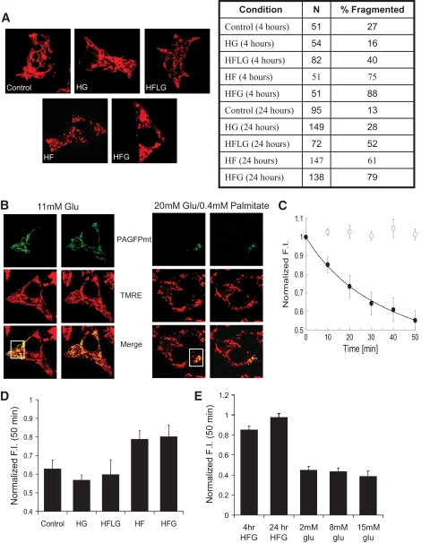
Culturing INS1 cells in media with high levels of fatty acids and glucose impairs mitochondrial morphology and dynamics. A: Confocal images of INS1 cells stained with TMRE after 24 h in control (11 mmol/l glucose), HG (20 mmol/l glucose), HFLG (5 mmol/l glucose and 0.4 mmol/l palmitate), HF (11 mmol/l glucose and 0.4 mmol/l palmitate), and HFG (20 mmol/l glucose and 0.4 mmol/l palmitate) media. HF and HFG induce mitochondrial fragmentation within 4 h as opposed to incubation with control, HFLG, and HG media, which induce little or no effect on morphology. Cells possessing >50% fragmented mitochondria were considered fragmented. B: Assessment of mitochondrial fusion activity using the same approach as in Fig. 3. A group of mitochondria were labeled using a 2-photon laser (□, inset). Through fusion events, photoactivated GFP distributed itself throughout the mitochondrial network within 50 min in cells treated with control media. Redistribution is accompanied by dilution of the photoactivated form of PAGFPmt, revealed by the decreased PAGFPmt FI. In cells pretreated with HFG for 24 h (right panel), the activated PAGFPmt remained segregated in the mitochondria where it was initially photoactivated; dilution does not occur and PAGFP FI remains high. C: Quantitative summary of PAGFPmt dilution within the mitochondrial population after 24 h of HFG. ○, Cells exposed to HFG for 24 h; ●, cells incubated in normal growth media for the same amount of time. Average GFP dilution values were fitted (R = 0.99) to a hyperbolic function yielding T50 of 10.1 min only for the normal media group. D: Mitochondrial fusion activity, measured by the ability to dilute PAGFPmt after 50 min. Histogram shows steady-state values of GFP FI obtained 50 min after photoactivation, when the PAGFPmt dilution reached equilibrium. HFG- and HF-treated cells show reduced fusion activity compared with control and HFLG- and HG-treated cells (P <0.05). E: A 4-h HFG treatment is sufficient to reduce the fusion activity of INS1 cell mitochondria to levels similar to those found with 24-h HFG treatment. Mitochondrial fusion activity is not affected by 30-min challenge with 8 and 15 mmol/l glucose. Glucose media was changed from 2 to 8 or 15 mmol/l 10 min prior to photoactivation. The plateau GFP FI level after 50 min is similar to that of the INS1 cells that remained in 2 mmol/l glucose. (A high-quality digital representation of this figure is available in the online issue.)

