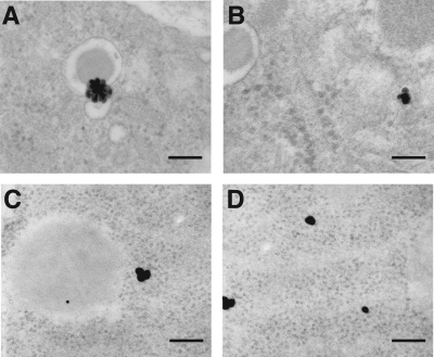
FIG. 4.
Ultrastructural analyses of exact intracellular localization of the SCAs after intravenous application in a rat. Transmission electron microscopy detected the β-cell–specific SCA B1 (A and B) and the α-cell–specific SCA A1 (C and D) in the endoplasmatic reticulum (A and C) and at the secretory granule membrane (B and D) of the respective target cells exclusively (n = 4 rats per group, 20–30 sections per rat). Scale bars, 90 nm.

