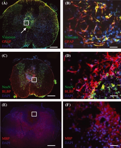
Figure 4.
Merged images of immunohistochemically stained transverse sections of injured spinal cord tissue taken from CsA-treated group at 3 weeks post-injury. (A,B) Vimentin (green) and GFAP (red) immunoreactive cells. The orange colour around the edge of the section shows that astrocytes contribute to the glia limitans. Some orange is also observed around the lesion. Expression of vimentin-positive, GFAP-negative staining around the central canal is obvious in (A). Region within white box in (A) is shown in (B). Arrow in (B) shows central canal of spinal cord. (C,D) BLBP (red) and NeuN (green) immunoreactive cells. Most BLBP positive astrocytes are located in region of glial scar, whereas NG2 positive cells are located within lesion. NeuN staining shows the dispersion of neuronal nuclei in the spinal cord section with an obvious concentration in the grey matter. (E,F) MBP (red) immunoreactive cells. MBP staining shows the dispersion of oligodendrocytes in the spinal cord section with obvious staining within the lesion which may relate to myelin debris that persisted following injury. Regions within white boxes in (A,C,E) are shown in (B,D,F), respectively. DAPI (blue) counterstain was used to label cell nuclei in all images. Scale bars: (A,C,E) 500 µm, (B,D,F) 30 µm.

