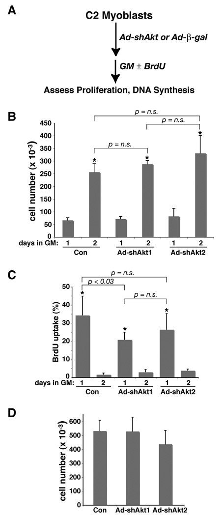
Figure 3. Knockdown of Akt1 or Akt2 does not inhibit C2 myoblast proliferation.
A. Experimental scheme. C2 myoblasts were infected at 25,000 cells per well (~5% of confluent density) with Ad-β-gal (control - Con), Ad-shAkt1, or Ad-shAkt2 (B), or at 50,000 cells per well (C, D), followed by incubation in growth medium (GM) for 1 – 3 days. B. Cells were counted at 1 and 2 days after viral infection (mean ± SD, n = 6; * - p < 0.0003 vs. day 1; other statistics are as indicated). C. BrdU incorporation into DNA was measured at 1 and 2 days after viral infection (mean ± SD, n = 6; * - p < 0.0003 vs. day 2; other statistics are as indicated). D. Cells were counted at 2 days after viral infection (mean ± SD, n = 6; p = n.s. for all pair-wise comparisons).

