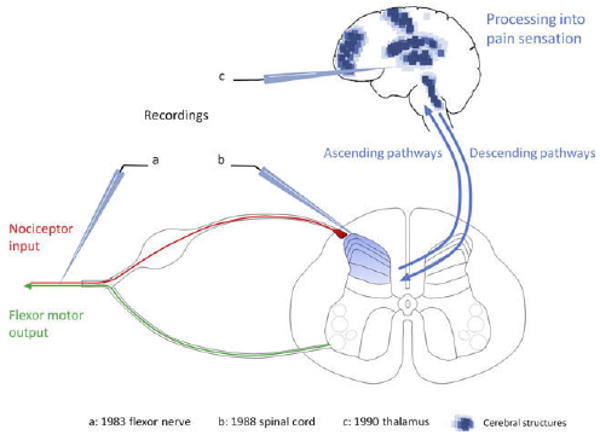
Figure 3.

Schematic representation of the structures exhibiting central sensitization. The first evidence for central sensitization was generated in 1983 by revealing injury-induced changes in the cutaneous receptive field properties of flexor motor neurons as an integrated measure of the functional plasticity in the spinal cord. A conditioning noxious stimulus resulted in long-lasting reductions in the threshold and an expansion of the receptive field of the motor neurons that was shown to be centrally generated. Essentially identical changes were then described in lamina I and V neurons in the dorsal horn of the spinal cord (b) as well as in spinal nucleus pars caudalis (Sp5c), thalamus (c), amygdala, and anterior cingulate cortex. Imaging techniques have revealed several brain structures in human subjects that exhibit changes compatible with central sensitization (blue dots).
