
Figure 3.
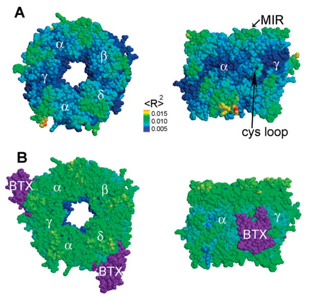
Motion of the extracellular domain of the AChR and its complex with αBTX. (A) Bottom and side views of the extracellular domain of AChR. The receptor twists, as the top and bottom rotate in opposite directions. (B) Bottom and side views of the extracellular domain of AChR in complex with αBTX. The receptor vibrates as a single unit and does not twist. The motion amplitude is presented through color coding. Blue, green, and orange are indicative of little, medium, and large motions respectively. For clarity, the αBTX molecules are colored purple. Indicated are α, δ, β, and γ subunits of the receptor, αBTX (BTX), the Cys loop, and the MIRs.