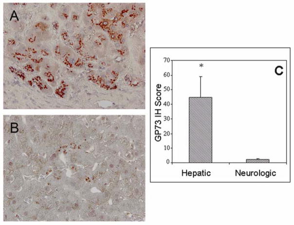
Fig. 2.
Hepatocyte expression of GP73 in WD with predominant hepatologic or neurologic manifestation. GP73 IH staining in livers from patients with primary hepatologic (A) or neurologic (B) presentations of WD. In (A), robust immunoreactivity is present in hepatocytes, with a pericanalicular pattern. Little or no immunoreactivity is present in fibrous septa and in non-hepatocyte cell types within the lobules. In (B) minimal or no immunoreactivity is present in hepatocytes. Individual perisinusoidal cells show GP73 immunoreactivity. Original Magnification 20×. (C) GP73 IH staining intensity scores (mean ± standard error) of hepatocyte GP73 expression in WD patients with primary liver and neurological involvement. Significance levels; *, p<0.05.

