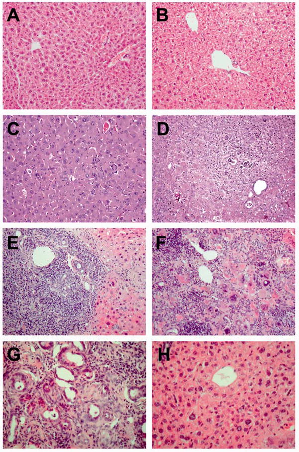
Fig. 4.

Histology of Atp7b−/− mouse livers. H&E stained liver sections from a 6 week-old wild-type female (A), a 32 week old wild-type female (B), a 6 week-old Atp7b−/− female showing normal histology (C), a 20 week-old Atp7b−/− female showing hepatitis and a bile duct lesion (D), a 32 week-old Atp7b−/− female showing severe hepatitis and a microabcess (E), a 46 week-old Atp7b−/− female showing severe necrosis and hepatitis (F), a tumor from a 60 week-old Atp7b−/− female showing neoplastic bile duct proliferation (G), and regenerating liver tissue with normal histology (H) from the same animal as (G). Original magnification 100× (A, B), 200× (C–G).
