
Figure 2.
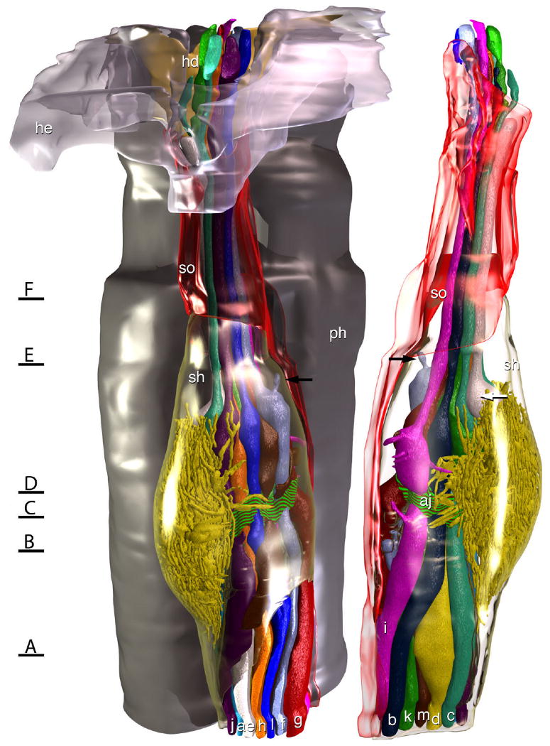
Model of the amphid of Acrobeles complexus. In the rendering on the left the left amphid is viewed from the left side of the animal, and in the rendering on the right the same amphid is viewed from the opposite side. The amphid socket cell (so) is rendered transparent red and the sheath cell (sh) is rendered transparent green. For reference, on the left side a portion of the HYP-E epidermal syncytium (he) is rendered transparent gray and a portion of the HYP-D epidermal syncytium (hd) is rendered solid yellow, showing where they form the amphid opening to the outside environment. Behind the amphid, a portion of the pharynx is rendered in gray. A belt of adherens junctions at the point where dendritic processes enter the sensory channel (aj) is indicated by a red and green striped texture. A small accessory process of ASF (black arrows) terminates in a pocket of the sheath cell, rather than in the sensory channel of the amphid. The bulbous swelling of ASE (white arrow) rests between the two cilia of ADC. Black lines with letters indicate the approximate region of the amphid from which the images in Figure 1 were obtained.