
Figure 3.
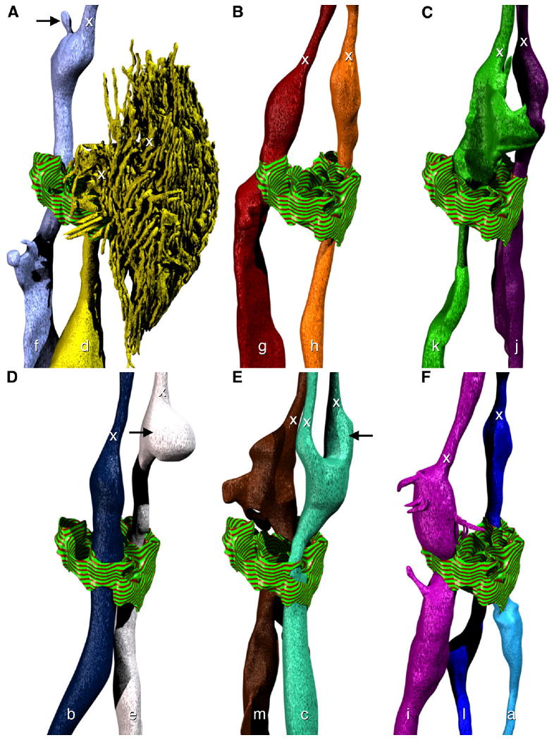
Model of the morphology of individual amphid dendrites. Adherens junctions at the opening in the sheath cell to the sensory channel are indicated with a striped green and red texture. The positions of transition zones (x) are indicated for all dendrites. The perspective is identical to the right-hand side of Figure 2. A: The neurites ASF (f) and ADF (d). Note the small projection (black arrow) near the transition zone. B: The neurites ASG (g) and ASH (h). C: The neurites ASK (k) and ASJ (j). D: The neurites ASB (b) and ASE (e). The bulbous swelling (black arrow) of ASE rests between the two cilia of ADC. E: The neurites ASM (m) and ADC (c). The cilium of ADC that is further from the pharynx has a more robust swelling near the transition zone (black arrow). F: The neurites ASI (i), ASL (l), and ASA (a).