
Figure 4.
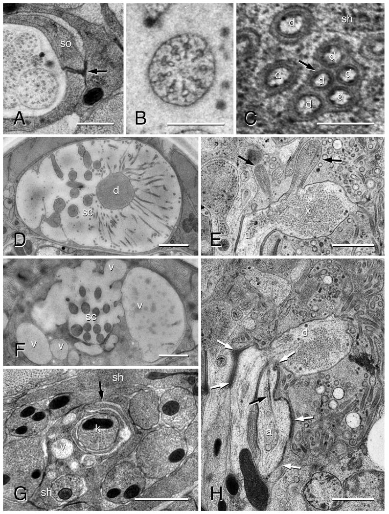
Transmission electron micrographs of an amphid in an adult female of Acrobeles complexus. A: Anterior region of amphid sensory channel showing the self-junction (black arrow) of the socket cell (so). B: Transition zone of an amphid sensory cilium. C: Distinctive cell membranes (black arrow) of the AFD microvilli (d) shown adjacent to the sheath cell (sh) membrane. D: Amphid in the region of the AFD (d) microvilli. The amphid sensory channel (sc) in this individual is filled with a secretion from the sheath cell. E: Longitudinal image through the amphid finger cell (d) showing two sensory cilia (black arrows) and granular material in the cytosol of the finger cell bulb (x). F: The same individual as in D, shown in the anterior region of the sensory channel (sc) where large secretory vesicles (v) merge with the sensory channel. G: Lamellar projections (black arrow) of the sheath cell (sh) wrapping around the sensory dendrite ASK (k) just posterior to where it enters the sheath cell sensory channel. Note the numerous vesicles (v) in this region. Scale bar = 1 μm. H: Longitudinal section through the amphid. The partially formed cilium of ASA (a) is shown terminating adjacent to the AFD cell (d). The rootlet is indicated (black arrow). Note the greater surface area of adherens junctions (white arrows) along ASA as compared to the other sensory dendrites. Scale bars = 0.5 μm in A; 0.25 μm in B,C; 1 μm in D–H.