
Figure 5.
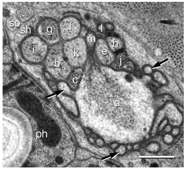
Transmission electron micrograph of an amphid in the J1 of Acrobeles complexus. The sensory dendrites ASB (b), ADC (c), AFD (d), ASE (e), ASF (f), ASG (g), ASH (h), ASI (i), ASJ (j), ASK (k), ASL (l), and ASM (m) are shown at the entrance to the sheath cell sensory channel. Also indicated are the sheath cell (sh), socket cell (so), and the pharynx (ph). The AFD cell is surrounded by microvilli, three of them are indicated (black arrows). Scale bar = 0.5 μm.