
Figure 6.
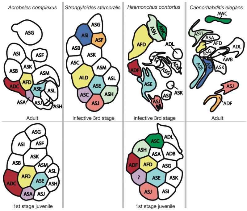
Arrangement of amphid dendrites at the posterior entrance into the sheath cell. The bottom row is from J1 while the top row is from the oldest life stage for which there is data. The right side of each image is adjacent to the pharynx, and the top is dorsal. Letters correspond to the name of dendrites according to current nomenclature. Cells that are colored the same represent stronger hypotheses of homology that are based on multiple types of similarity, with dendrites of the same color being presumably homologous. Types of similarity considered include number of cilia, results of laser ablation studies that imply related function, and relative position at the proximal end of the amphid. Data for Strongyloides stercoralis are redrawn from Ashton et al. (1995). Data for Haemonchus contortus are redrawn from Li et al. (2000a, 2001). Data for C. elegans are redrawn from Ward et al. (1975).