
Figure 4.
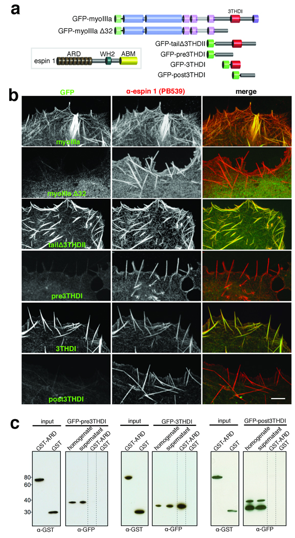
Myosin IIIa interacts with espin 1 through its 3THDI domain. (a) Schematic representation of the espin 1 and myosin IIIa constructs analyzed in this figure. Legend: ABM, actin binding module; WH2, Wiskott-Aldrich homology domain 2; GFP-myoIIIa Δ32, myosin IIIa lacking exon 32 which causes a frame shift rendering the protein without the 3THDI and 3THDII domains. (b) Co-expression of untagged espin 1 shows that GFP-myoIIIa, GFP-tailΔ3THDII, and GFP-3THDI (green) colocalize with espin 1 (red) along actin filament bundles. In contrast, GFP-myoIIIa Δ32, GFP-pre-, and post3THDI are dispersed in the cytoplasm, despite the presence of espin 1 bundles. Scale bar, 5 µm. (c) Western blots of GST pull-downs confirm that the 3THDI region of myosin IIIa is necessary and sufficient for binding to espin 1 ARD, as pre3THDI and post3THDI show no binding to GST-ARD. Precipitates were detected using α-GST and α-GFP.