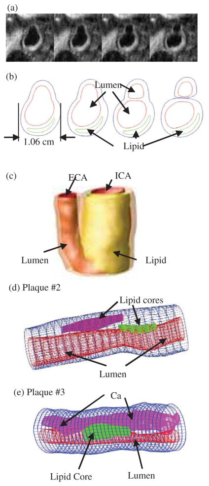
Figure 2.
In vivo 3D MRI images of a human carotid plaque and 3D reconstruction. (a) 4 MRI (T1W) slices (S2–S5) selected from a 24-slice set (S0–S23), slice spacing: 0.5mm. Each image shown here is cut from the whole-neck image; (b) segmented contour plots using CASCADE showing plaque components; (c) 3D geometry reconstructed from in vivo images; (d)–(e) two more plaque samples reconstructed from ex vivo MR images.

