Figure 1.
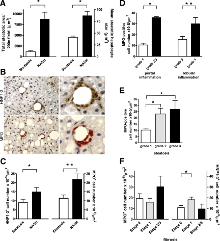
Increased MPO-positive cell number in NASH is associated with steatosis. A: The total amount of steatosis as well as the mean size of steatotic hepatocytes is significantly higher in subjects with NASH as compared with subjects with simple steatosis (*P < 0.001). B: Frequent organization of HNP1-3-positive neutrophils (red/brown, upper panel) and MPO-positive neutrophils (red/brown, lower panel) into crown-like structures surrounding steatotic hepatocytes (magnification, left panels, ×200, right panels, ×540). C: Subjects with NASH display more HNP1-3-positive neutrophils and more MPO-positive cells in the liver as compared with subjects with simple steatosis (*P = 0.02 and **P = 0.03, respectively). D: Higher grades of portal and lobular inflammation are associated with increased numbers of MPO-positive cells (*P < 0.01 and **P < 0.05, respectively). E: Significantly higher number of MPO-positive cells in subjects with grade 2 and grade 3 steatosis (*P < 0.05). F: The number of MPO-positive cells is not related to fibrosis stage in NASH as analyzed by one-way analysis of variance (*P = 0.75), whereas the number of HNP1-3-positive cells is higher in subjects with stage 1 fibrosis (*P = 0.05).

