Figure 3.
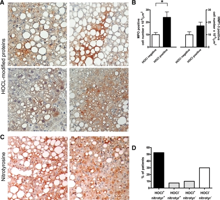
Accumulation of MPO-mediated HOCl-modified and nitrated proteins in NASH. A: Representative staining patterns for HOCl-modified proteins (red/brown) in livers of four different patients with NASH (magnification, ×200). More intense staining for HOCl-modified epitopes is frequently associated with steatotic hepatocytes. B: The number of hepatic MPO-positive cells is significantly higher in patients that show HOCl-modified protein staining (*P = 0.02). In contrast, HNP1-3-positive cell number is not significantly increased in this group (P = 0.14). C: Representative immunostainings of nitrotyrosine residues (red/brown) in livers of two patients with NASH (magnification, ×200). More intense staining is predominantly associated with steatotic hepatocytes. D: In most patients, HOCl-modified protein accumulation is coupled to nitrotyrosine accumulation.

