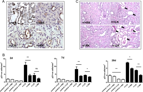
Figure 4.
The severity of tubular injury is increased in Pkd2 heterozygous mice compared with wild-type following IRI. A: Representative panels showing p21-positive tubular cells for both genotypes at 2 days post-IRI. Some background apical and cytoplasmic staining was seen with this antibody, but only nuclear positive cells (arrows) were counted. Magnification, ×400. B: The number of p21 nuclear positive tubular cells was quantified in age-matched control kidneys of both genotypes (white bars) and at 2, 7, and 28 days following IRI (black bars). There were significantly fewer p21-positive cells in heterozygous control kidneys compared with wild-type (significance shown on 28 days only for clarity). At all time points following reperfusion, tubular p21 expression was significantly lower in the heterozygous LK compared with wild-type LK. ∗P < 0.05, ∗∗P < 0.01. C: The panels show representative periodic acid-Schiff-stained kidney sections from the most severely affected animals of both genotypes (n = 6–7) with arrows indicating dilated tubules, some with cell debris and proteinaceous casts. Magnification, ×400. The mean percentage of necrotic tubules per section at 2 days post-IRI was greater in heterozygous mice but did not reach statistical significance.

