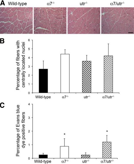
Figure 3.
α7/utr−/− mice exhibit a subtle muscle phenotype. A: H&E staining of triceps muscle from 5-week-old mice was used to examine muscle pathology. Wild-type, α7−/−, utr−/−, and α7/utr−/− contained few muscle fibers with centrally located nuclei. Scale bar = 50 μm. B: Quantitation of centrally located nuclei in triceps muscle of wild-type, α7−/−, utr−/−, and α7/utr−/− mice. Wild-type and utrophin null mice showed few centrally located nuclei. α7−/− and α7/utr−/− mice showed a slight increase in the percentage of myofibers with centrally located nuclei but these were not statistically different from wild-type controls (N = 6 mice/genotype). C: Evan’s blue dye uptake confirmed that sarcolemmal integrity was not further compromised in α7/utr−/− mice. Wild-type, α7−/−, and utr−/−, α7/utr−/− skeletal muscle showed fewer than 2% of muscle fibers that were Evan’s blue dye positive. (*P < 0.05, N = 5 mice/genotype).

