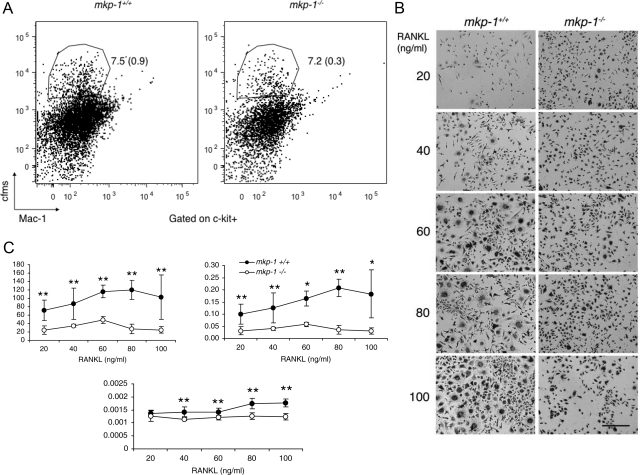
Figure 5.
Absence of MKP-1 reduces osteoclastogenesis in response to M-CSF and RANKL. A: Bone marrow-derived macrophages from 6-week-old MKP-1-deficient and wild-type mice were cultured in the presence of M-CSF (30 ng/ml) for 2 days before being subjected to flow cytometric analysis with antibodies directed against c-fms, Mac-1, and C-kit, as surface markers. Note that the absence of MKP-1 did not affect the number of osteoclast precursor cells. n = 3. B: Spleen-derived macrophages were cultured for 6 days in the presence of M-CSF (20 ng/ml) and increasing concentrations of RANKL. Scale bar = 100 μm. Cells were stained for the osteoclast marker tartrate-resistant acid phosphates to record their number per well, the surface they occupy, with calculation of average cell size (C). n = 5. Data are means ± SD. *P < 0.05 and **P < 0.01.

