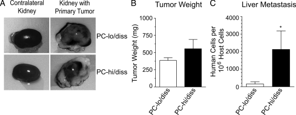
Figure 2.
Dissemination of PC-3 variants in the renal capsule spontaneous metastasis model. A: Selected PC-lo/diss and PC-hi/diss cells were grafted under the kidney capsule of nu/nu mice. Large primary tumors developed on the kidneys and almost completely engulfed them within 4 to 5 weeks post implantation (right panels). For comparison, contralateral kidneys are shown on the left. B: The net weight of individual primary PC-lo/diss and PC-hi/diss tumors was estimated by subtracting the weight of the contralateral kidney. Data are means ± SEM from a representative experiment using 4 mice per variant. C: Metastasis levels in the livers of mice bearing PC-lo/diss (open bars) and PC-hi/diss (closed bars), quantified by Alu-qPCR. *P < 0.05 in one-tailed Mann-Whitney test.

