Figure 1.
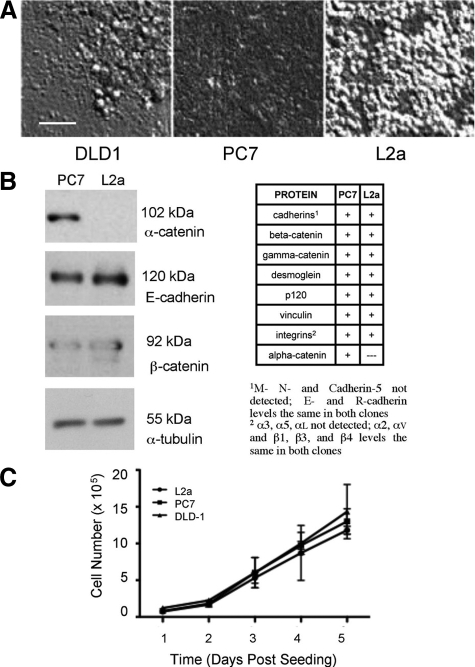
Morphological differences between clones of DLD-1 colorectal carcinoma cells. A: DLD-1 cells display variable cell-cell and cell-matrix adhesion characteristics, as revealed by differential interference contrast microscopy. Clone PC7 forms a tightly compacted monolayer with indistinct cell-cell boundaries, whereas clone L2a cells display a “rounding” phenotype, with a less compact layer on the culture dish, covered by patches of multilayered clusters of weakly adherent cells. DLD-1 parental cultures are a mixture of both morphologies. B: Expression of proteins associated with cell-cell and cell-matrix adhesion was evaluated by Western blotting. In all of the proteins examined, the only quantitative difference between PC7 and L2a was that the later clone was negative for the adherence junction protein α-catenin. C: Despite the obvious variation in cell morphology, no significant differences were seen in cell proliferation between DLD-1 parent cells or either clone. Scale bar = 50 μm.

