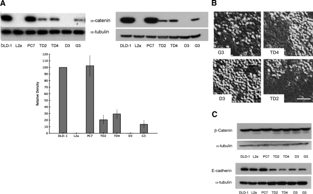
Figure 3.
α-Catenin levels in L2a clones transfected with expression construct. A: Western blots of cell lysates from DLD-1 parent cells, PC7, L2a, and α-catenin expression-derived clones (TD2, TD4, D3, and G3). Blots were probed with antibodies recognizing the C-terminal (left) and N-terminal (right) domains of α-catenin protein. L2a cells lack detectable α-catenin. Re-expression of α-catenin was successful in clones TD2, TD4, and G3 but not clone D3. The graph shows densitometric analysis of α-catenin protein levels (normalized to tubulin signal) expressed as a percentage of DLD-1 parent cell expression. L2a and D3 clones lines have no detectable α-catenin expression (*P < 0.001). B: Differential interference contrast microscopy of transfected clones illustrating relative amounts of “rounded” (α-catenin-null) cells. As expected from the protein levels seen in A, all clones except D3 have normalized regions of monolayer consisting of tightly adherent cells. Scale bar = 50 μm. C: Western blots for β-catenin and E-cadherin in DLD-1 derivatives and α-catenin re-expression clones demonstrate no significant loss of other elements of the adhesion junction complex.

