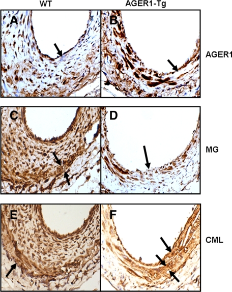
Figure 6.
AGER1 expression and the location and amount of AGEs in AGER1-tg and wild-type (WT) mice. Analysis of femoral artery sections by immunohistochemistry after a high-fat diet (8 weeks) and wire-induced injury (4 weeks) in AGER1-tg mice (n = 14) and wild-type mice (n = 13). Representative sections of injured vessels from wild-type (A, C, and E) and AGER1-tg mice (B, D, and F), stained with anti-AGER1 (A and B), anti-MG (C and D), and anti-CML (E and F) antibodies, as described. Arrows indicate cellular localization of AGER1 (A and B), the thinned media, compared with thickened and cellular intima in wild-type mice (C) and to the normal intima of AGER1-tg mice (D); and an area of fibrosis (F). A: Many cells in the hyperplastic intima do not contain AGER1 (arrow). B: The media contains scattered areas of fibrosis (arrow). C: The media (arrows) underlying the hyperplastic intima is thinned; intracellular MG staining is present in all cell layers. D: Cells in the fibrotic areas of the media are smaller, but contain MG intracellularly. E: CML is present in the cytoplasm of cells of all three layers, and in the focally dense extracellular matrix of the media (arrow). F: CML is present in the extracellular matrix of the occasional plaques (arrow) and in the fibrotic areas of the media (double arrows).

