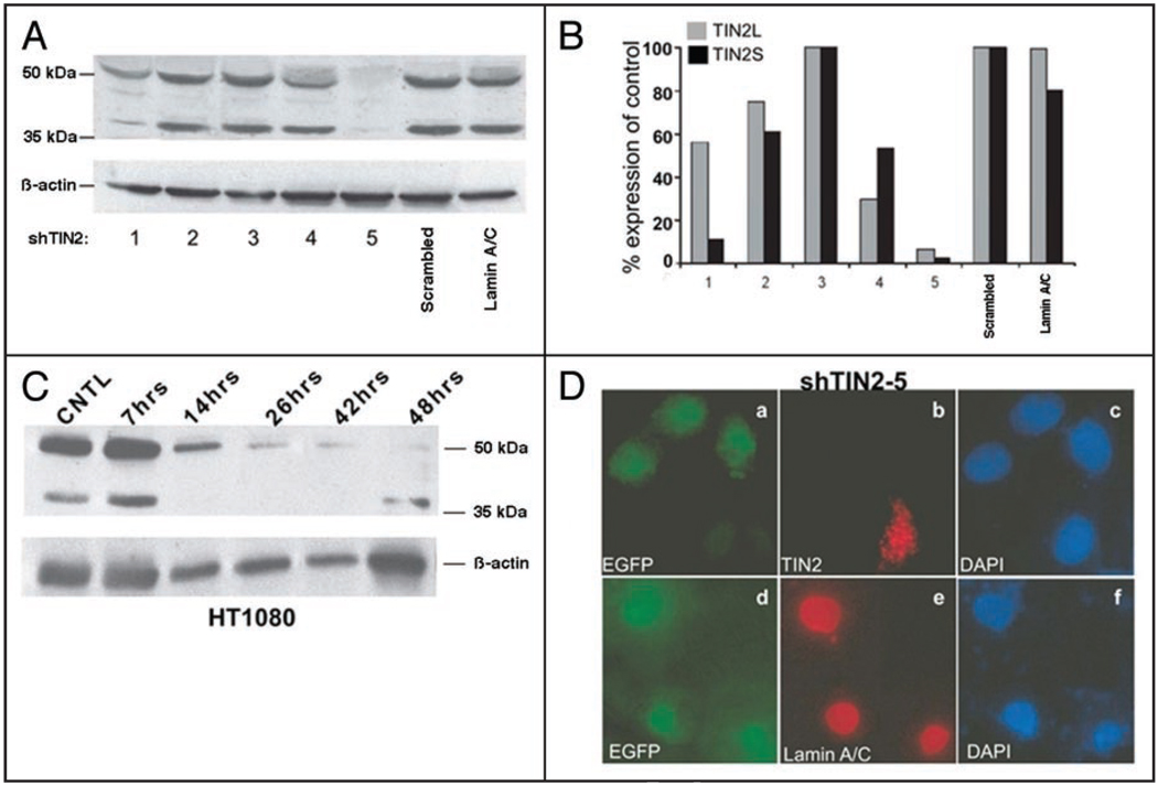
Figure 2.
RNAi reduces both TIN2S and TIN2L. (A) Western analyses of hTIN2 proteins in cells expressing different shRNA constructs. HT1080 cells were transiently transfected with vectors expressing different shRNAs designed to target hTIN2 (shTIN2-1 to 5). The cells were lysed in 2X Laemmli buffer 36 h later and analyzed for hTIN2 proteins and β-actin (protein loading control). Controls for non-specific shRNA effects include a scrambled shRNA sequence and a pre-validated lamin A/C shRNA. (B) Quantification of the western blot shown in (A). The signals were quantified by densitometry and the hTIN2 signals were normalized to the β-actin signals. The normalized signals are displayed as a percentage of signals generated by the scrambled shRNA control. (C) Time course of hTIN2 knockdown. HT1080 cells were transiently transfected with the shRNA 5 vector. At the indicated intervals, the cells were lysed and analyzed for hTIN2 proteins and β-actin by western blotting. (D) Immunofluorescence analysis of hTIN2 depletion by shRNA. HT1080 cells were transfected with the vector expressing shTIN2-5. Transfected cells were identified by EGFP expression (green; a and d); the nuclei of all cells were identified by DAPI staining (blue; c and f). The cells were immunostained for hTIN2 (red; b) or lamin A/C (red; e). Cells were visualized by fluorescence microscopy.

