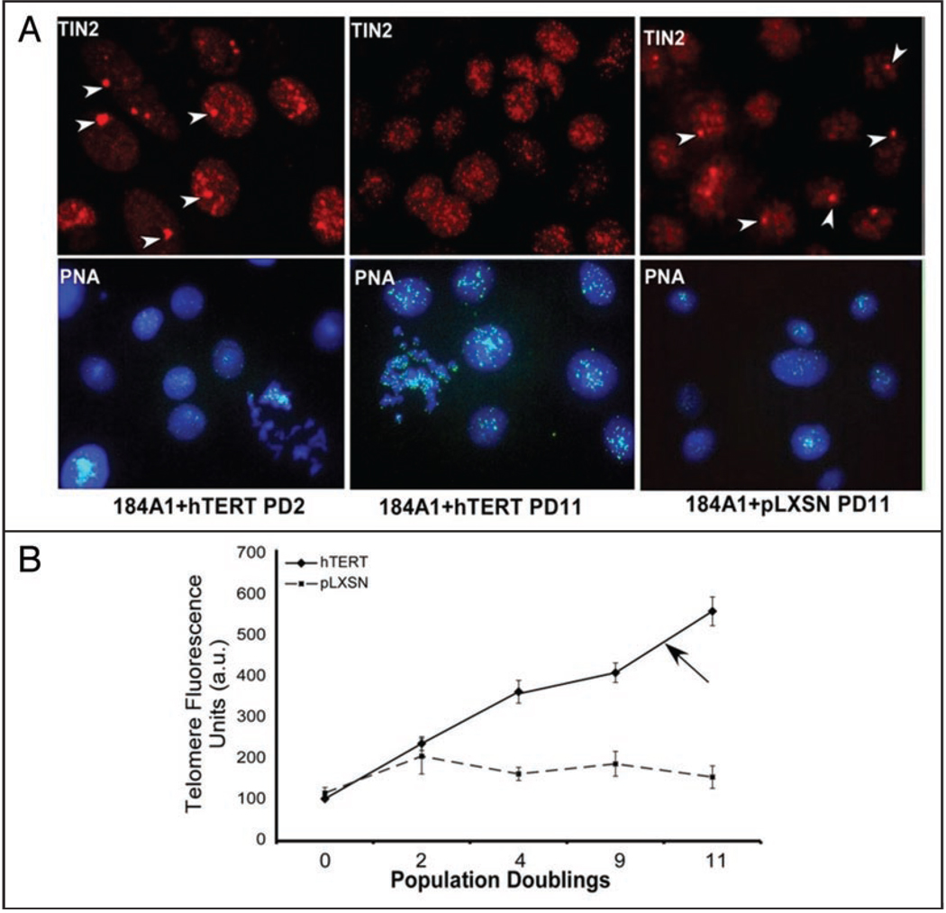
Figure 6.
hTIN2S, but not hTIN2L, localization depends on telomere length. (A) 184A1 HMECs were infected with a control retrovirus (pLXSN) or retrovirus expressing the catalytic subunit of telomerase (hTERT). The cells were immunostained for TIN2 (red) 2 and 11 population doublings (PD) after infection. Arrows indicate non-telomeric TIN2 domains. Parallel cultures were analyzed for telomere length using qFISH (green), as described in Materials and Methods. Nuclei were counterstained with DAPI (blue). (B) Quantification of average fluorescence from qFISH analyses. Fluorescence intensities from >300 cells were measured. The results are expressed in arbitrary units (a.u.) relative the fluorescence intensities of uninfected cells (PD 0). Arrow indicates the telomere length at which non-telomeric TIN2 domains are no longer visible.

