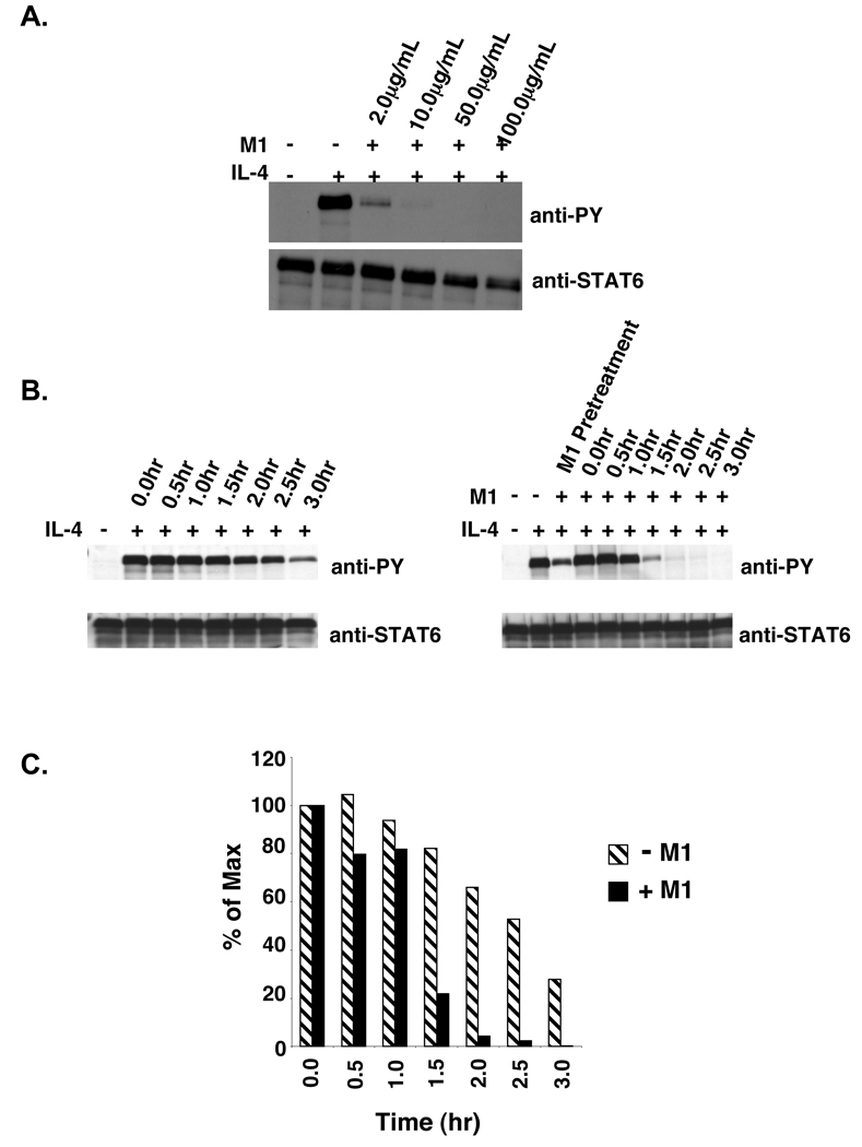
Figure 5. M1 blocking of murine IL-4 stimulated V50-IL-4Rα U937 clones.
A. V50-IL-4Rα U937 cells were treated with various concentrations of the M1 anti-mIL-4Rα blocking antibody (2.0 µg/mL, 10.0 µg/mL, 50.0 µg/mL, and 100.0 µg/mL) for 60 minutes. The cells were then stimulated with 10 ng/mL murine IL-4 for 15 minutes. The cells were lysed and STAT6 was immunoprecipitated and subjected to western blot analysis using an anti-phosphotyrosine antibody. The blot was stripped and reprobed with an anti-STAT6 antibody to detect STAT6. B. V50-IL-4Rα U937 clone #5 was stimulated in either the absence or presence of 10 ng/mL murine IL-4 for 15 minutes. Post stimulation, the IL-4 was washed out and the cells were re-cultured in the presence or absence of 10 µg/mL of the anti-murine IL-4Rα blocking antibody M1. The cells were lysed at the indicated time points and STAT6 was immunoprecipitated and subjected to western blot analysis using an anti-phosphotyrosine antibody. The blots were stripped and reprobed with an anti-STAT6 antibody to detect STAT6. C. The film shown in Panel B was scanned and NIH-Image 1.63 was used to determine the densities of the bands developed on the western blots. The ratio of phosphorylated STAT6 to total STAT6 was calculated and the percent max was determined and graphed.

