
Figure 1. Structures of A) Inhibitor 1 (GRL-98065), B) Darunavir (TMC-114), and C) PR dimer indicating location of mutations.


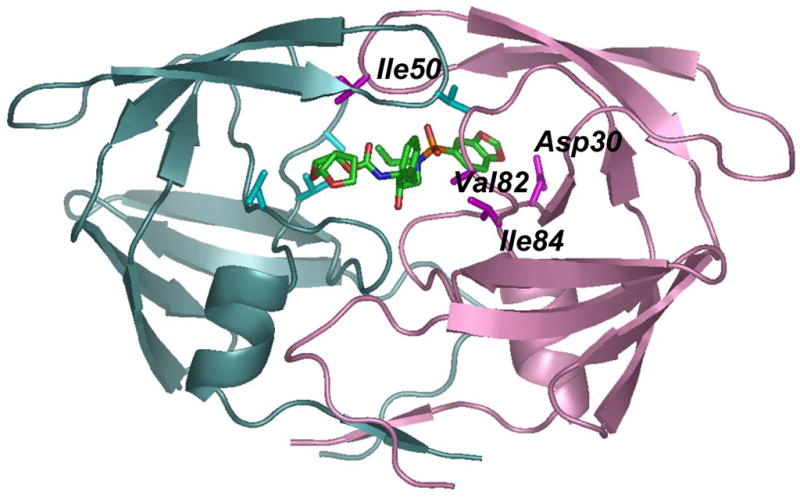
The backbone of one subunit is shown as pink ribbons with the sites of mutation (D30N, I50V, V82A and I84V) indicated by magenta side chains, while the other subunit is colored in light cyan with mutation sites in darker cyan. Only one subunit is labeled. Inhibitor 1 is colored by atom type.