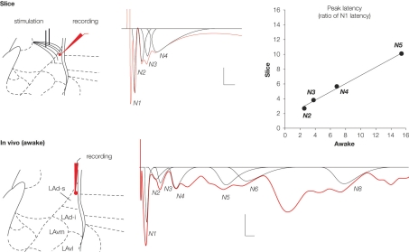
Figure 3.
Field potentials (in red) evoked in the lateral amygdala (LA) by stimulation of auditory afferents (internal capsula in vitro, auditory thalamus in vivo) showed multiple negative potentials both in vitro (slice) and in vivo (awake rat). Scale: 0.1 mV, 10 ms. These waves represent a monosynaptic response (N1) followed by polysynaptic activity. We developed a method for fitting and extracting peaks that represent reliable synchronized activity (in black). Five and nine peaks were isolated in the slice and the awake preparation, respectively. Note that not all peaks are present in a given sample (e g. here N5 is missing for the in vitro example, or N7 for the in vivo example). To enable comparison between the two preparations, latencies of each of the isolated peaks were normalized with respect to the latency of the first monosynaptic peak (N1). This transformation showed a high correlation between the two preparations (r2 = 0.994; P < 0.005), suggesting similar intra-network rhythmicity in both preparations, and therefore similar network behavior.

