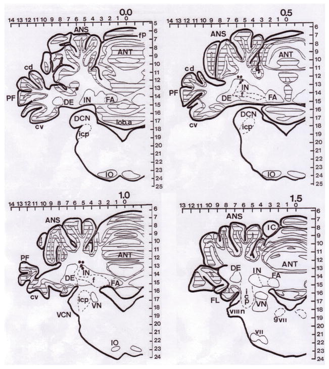
Figure 2.
Histological reconstruction of marking interpositus nucleus (IP) lesions on sections from the short-term study. ANS = ansiform lobule; ANT = anterior lobe; PF = paraflocculus; IN = interpositus nucleus; f = fibers; DE = dentate nucleus; FA = fastigial nucleus; cv = ventral crus; icp = inferior cerebellar peduncle; IO = inferior olive; VN = vestibular nucleus; VCN = ventral cochlear nucleus. Each dot represents the tip of infusion cannular seen in each brain slice. Due to the localization technique, the some lesions could be found in more than one slice.

