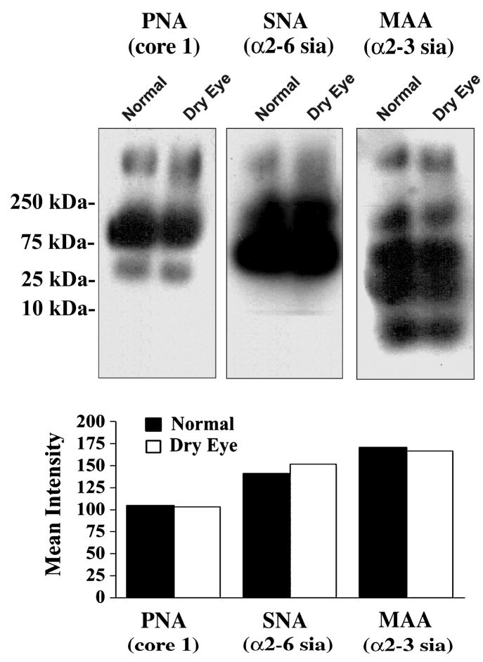
Figure 4.
Lectin binding to glycoproteins in tear fluid of normal subjects and dry eye patients. Samples containing 7.5 μg of total protein were separated by 1% agarose gel electrophoresis and blotted to nitrocellulose membranes. Glycan content was assessed using biotin-labeled PNA to the mucin-associated T-antigen epitope, SNA to terminal α2-6 sialic acid, and MAA to terminal α2-3 sialic acid. The position of the molecular weight markers is indicated to the left. As determined by densitometry (lower panel), no differences in lectin binding were detected between tear samples from normal subjects and dry eye patients.

