
Figure 1. Neural canal architecture, demonstrating the neural canal opening (NCO) and Border Tissue within a histologic section (upper) and a co-localized spectral domain OCT B-scan.
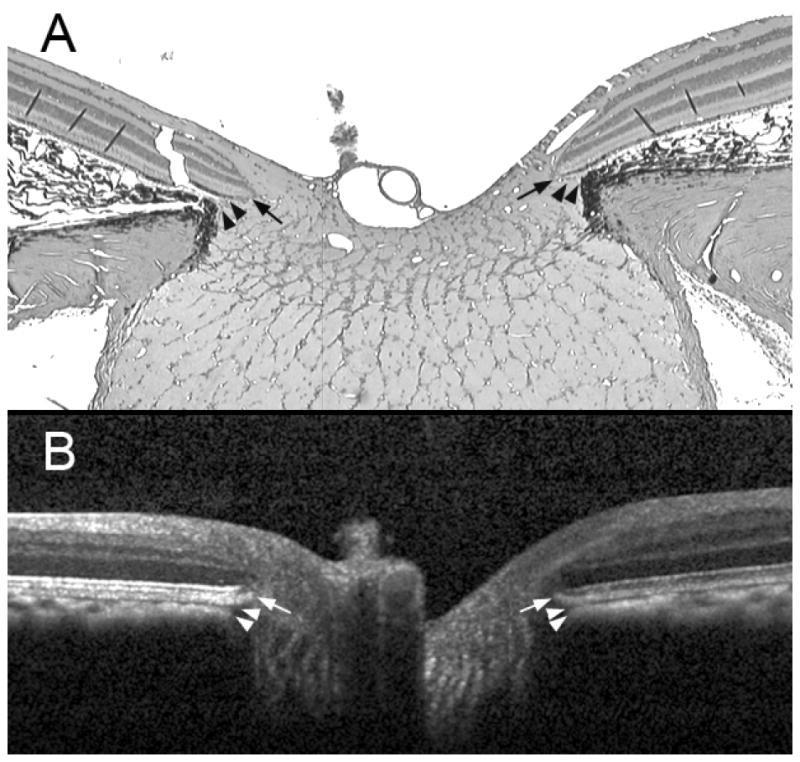
A representative serial histologic section (hematoxylin and eosin stain, × 2.5 magnification) through the optic nerve head of a normal monkey eye (not part of this report), perfusion fixed at an IOP of 10 mmHg (panel A) compared to an equivalent B-scan acquired in vivo from the same optic nerve head at an IOP of 10 mmHg (panel B). The arrows show the position of the NCO, which in the histologic section (black arrows) is at the termination of Bruch’s Membrane either side of the neural canal and in the B-scan (white arrows) is at the termination of the retinal pigment epithelium/Bruch’s Membrane signal either side of the neural canal. The arrowheads show the position of Border Tissue, which in the histologic section (black arrowheads) is an extension of the scleral connective tissue, enclosing the choroid to meet Bruch’s Membrane; in the B-scan (white arrowheads) the Border Tissue is identified as the junction of the choroidal signal with the neural canal. The neural canal extends from the NCO until the point where the optic nerve exits the globe, passing through a choroidal component (bound by the Border Tissue) followed by a scleral component.