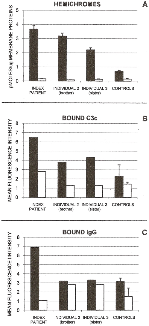
Figure 1. Membrane-bound hemichromes, complement C3c fragment, and autologous IgG in GR-deficient and GR-sufficient RBCs.
Shown are membrane-bound hemichromes (panel A), complement C3c fragment (panel B) and autologous IgG (panel C) in ring-infected and non-infected GR-deficient RBCs from the index patient, brother (individual 2) and sister (individual 3) of index patient, and from normal GR-sufficient controls. Black bars indicate ring-infected RBCs; open bars, non-infected RBCs. Hemichromes are expressed as pmoles/µg membrane protein. Mean values of index patient, individuals 2 and 3, and normal controls (mean±SD, n = 2–4) are shown. Hemichromes were significantly higher (p<0.02) in the ring-infected RBCs of the index patient and individuals 2 and 3 compared to ring-infected RBCs of normal controls. Complement C3c fragment and autologous IgG are expressed as Mean Fluorescence Intensity. Representative data of GR-deficient individuals and mean values of normal controls (mean±SD, n = 2–3). For experimental details see Materials and Methods.

