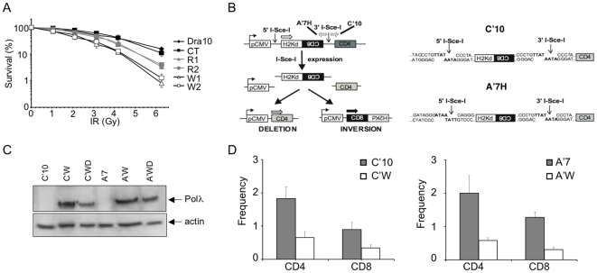
Figure 4. Defective NHEJ in cells expressing the polymorphic W438 hPolλ variant.
(A) IR sensitivity of the DRA10 cell lines expressing the different forms of hPolλ described in Figure 3. (B) Substrate used to measure NHEJ (left panel) and representation of the sequences of the I-Sce-I restriction sites in the C′10 and A′7 cell lines (right panel). (C) Expression of the different forms (active: C′W, A′W; inactive: C′WD, A′WD) of W438 hPolλ in C′10 and A′7 analyzed by Western blot. (D) Evaluation of the deletion (CD4) and inversion (CD8) events in C′10 (left panel) and A′7 cells (right panel) expressing the empty vector (white bars) and the indicated W438 variant of hPolλ (grey bars). Results are the mean+/−SD of 3 independent experiments, *represent significant statistical difference (P<0.05), **represent significant statistical difference (P<0.005).

