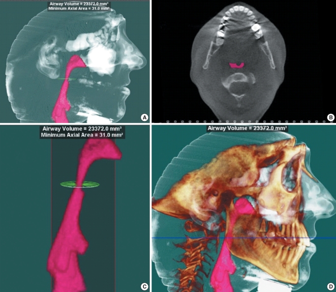
Fig. 1.
(A) is a 3-D CT taken awake and supine with software reconstruction specifically to assess characteristics of the airway. (B) is an axial section showing the minimum cross sectional area (MCSA) of the pharyngeal airway which measures 31.0 mm3. This is a significant narrowing at that level. (C) has a total airway volume of 23,372.2 mm3 from the inlet to the outlet outlined in pink. (D) is a reconstruction of the facial skeleton along with an outline of soft tissues. This allows an exposure of the airway that is not seen in traditional radiographs.

