Figure 7.
EIN3 Binds the SID2 Promoter.
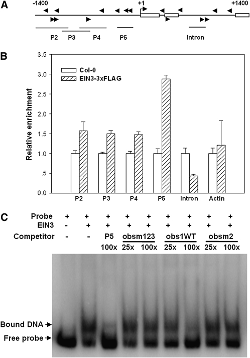
(A) Schematic diagram of potential EIN3 binding sites (arrows) and DNA fragments used for ChIP experiments. Shown are 1.4-kb upstream sequence and part of the coding sequence for SID2. Boxes are exons, and the translational start site (ATG) is shown at position +1.
(B) Enrichment of the indicated DNA fragments following ChIP using anti-FLAG antibody. Chromatin from wild-type and transgenic plants expressing EIN3-3×FLAG was immunoprecipitated with an anti-FLAG antibody, and the presence of the indicated DNA in the immune complex was determined by quantitative real-time PCR. The amounts of DNA amplified from the EIN3-3×FLAG seedlings were normalized to that from Col-0 plants. The Actin promoter fragment was used as a negative control. The experiment was repeated three times with similar results.
(C) EMSA assay for EIN3-P5 DNA fragment binding in vitro. Radiolabeled P5 DNA fragment was incubated with GST-EIN3 protein, and the free and bound DNA (arrows) were separated in an acrylamide gel. Where indicated, the P5 fragment and oligonucleotide primers WTobs1 (5′-AACGATGTACCTGGTCGTATT-3′), obsm2 (5′-GTACATTTACCTGGACCGTGA-3′), and obsm123 (5′-GTACCTTTCCCTGGACCGTGA-3′) were used as competitor DNA.

