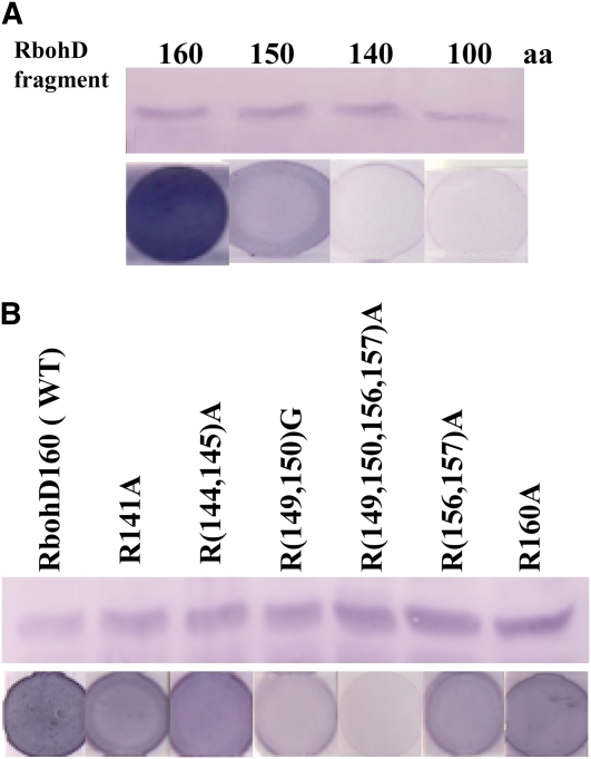
Figure 7.
Identification of PA Binding Sites in RbohD.
(A) Binding of RbohD fragments to PA. Top panel: immunoblotting of N-terminal fragments (1 to 100, 140, 150, and 160 amino acids, respectively) expressed in E. coli. Bottom panel: binding of protein fragments to PA on filters.
(B) Expression of mutated RbohD fragments and detection of PA binding. Top panel: immunoblotting of wild-type and mutated RbohD160 fragments expressed in E. coli. Bottom panel: binding of RbohD160 fragments to PA on filters using the same protein as in the top panel.
[See online article for color version of this figure.]

