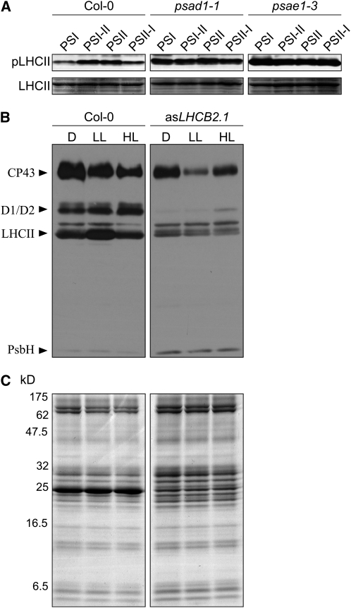
Figure 7.
Phosphorylation Patterns in Wild-Type (Col-0) and Mutants (psad1-1, psae1-3, and asLHCB2.1) Acclimated to the Conditions Used to Monitor LTR or State Transitions.
(A) Phosphorylated LHCII (Pi-LHCII; top panels) was detected by immunoblot analysis using a phosphothreonine-specific antibody in wild-type and mutant (psad1-1 and psae1-3) plants adapted to PSI or PSII light or shifted between the two light regimes (PSI-II; PSII-I). Portions of Coomassie-stained SDS-PAGE gels, identical to those used for blotting and corresponding to the LHCII migration region (bottom panels), were used to control for equal loading. Results of one of three independent experiments are shown.
(B) Thylakoid protein phosphorylation in the wild type (Col-0) and asLHCB2.1 plants acclimated to dark (D; 16 h), low light (LL; 80 μmol m−2 s−1, 3 h), and high light (HL; 800 μmol m−2 s−1, 3 h) conditions. Phosphorylated thylakoid proteins were detected by immunoblot analysis using a phosphothreonine-specific antibody. Results of one of three independent experiments are shown.
(C) Coomassie blue–stained SDS-PAGE gels, identical to those used for blotting shown in (B), were used to control for equal loading.

