Figure 2.
Infection Phenotypes of C. orbiculare ssd1 Mutant and Defense Responses on N. benthamiana.
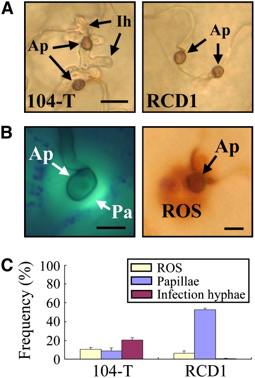
(A) Cytology of appressorial penetration by the wild-type 104-T and ssd1 mutant RCD1. The upper leaves were droplet-inoculated with conidial suspension (5 × 10⁵ conidia/mL) and observed at 72 HAI. Ap, appressorium; Ih, intracellular infection hyphae. Bar = 10 μm.
(B) Left panel: Callose papilla (Pa) formed under appressoria (Ap). The leaf was inoculated with 104-T, stained with aniline blue, and observed with epifluorescence microscopy at 72 HAI. Callose was detectable by its green fluorescence. Bar = 5 μm. Right panel: ROS accumulation under appressoria. The leaf inoculated with 104-T was stained with DAB and observed with light microscopy at 72 HAI. ROS was detectable as dark-brown staining. Bar = 5 μm.
(C) Quantification of papilla formation and ROS accumulation at sites of attempted penetration by C. orbiculare appressoria. At 72 HAI, leaves inoculated with the wild-type 104-T and ssd1 mutant RCD1 were stained with aniline blue to detect callose or with DAB to detect ROS accumulation. At least 200 appressoria were counted for each fungal strain, and standard errors were calculated from three replicate experiments.
[See online article for color version of this figure.]

