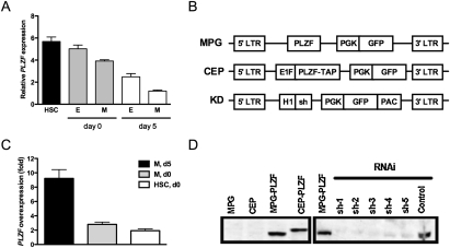
Figure 1.
PLZF expression and viral vector design. (A) Expression of PLZF mRNA in human hematopoietic subsets, before and after culture, was quantified by qPCR. Hematopoietic subsets were isolated by FACS-sorting from fresh Lin− CB (day 0) or after 5 d in serum-free culture (day 5) based on the following phenotypes: HSCs, CD34+ CD38−, erythroid progenitors (E), CD34+ CD38+ CD71+, myeloid progenitors (M), and CD34+ CD38+ CD71−. (B) Schematic representation of viral vectors used to overexpress (MPG, retroviral; CEP, lentiviral) or silence human PLZF (shPLZF). (C) Fold overexpression of PLZF in myeloid progenitors transduced with MPG-PLZF and cultured for 5 d, compared with same-day control cells (M, d5), or freshly sorted myeloid progenitors (M, d0) or HSCs (HSC, d0) from the same CB. (D, left panel) Western blot analysis of total protein extracts from 293T cells transduced with control or MPG-PLZF viruses. (Right panel) Silencing vectors (shPLZF1–3) were transduced into 293T cells stably expressing PLZF. Equal total protein was loaded in all lanes and the blots were probed with an anti-PLZF antibody.

