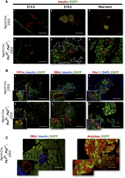
Figure 2.
Presenilin-deficient pancreatic progenitors increase in numbers late during embryonic development. (A) Immunostaining for insulin (red) and EGFP (green) at E14.0 (left) and E18.5 (middle) and in the newborn (right) in control (Ngn3-Cre; Z/EG) (top) and presenilin-deficient (Ngn3-Cre; Ps1f/−; Ps2−/−; Z/EG) (bottom) mice. EGFP-positive cells are found outside of the endocrine compartment in Presenilin mice throughout development and expand around birth. (B) Coimmunostaining for Ptf1a (red), insulin (blue), and EGFP (green) (middle); DBA (red), insulin (blue), and EGFP (green) (right panel); or Pdx1 (red) DAPI (blue), and EGFP (green) (left panel) in sections of E18.5 control (Ngn3-Cre; Z/EG) (top) and Presenilin-deficient (Ngn3-Cre; Ps1f/−; Ps2−/−; Z/EG) (bottom) mice. Pslow, nonendocrine cells still express Pdx1, Ptf1a, and DBA at E18.5. (C) Coimmunostaining for DBA (red), Insulin (blue), and EGFP (green) (left panel), or Amylase (red) and EGFP (green) (right panel). Pslow cells exclusively express Amylase in the adult. Normal, EGFP-negative acinar cells form clusters n the periphery of the tissue. Bars, 50 μm.

