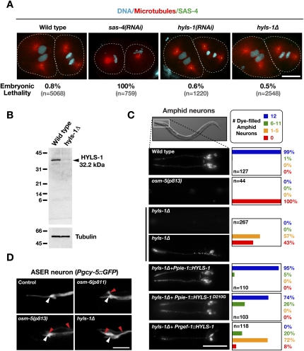
Figure 3.
HYLS-1 is dispensable for centriole assembly and centrosome function, but is essential for ciliogenesis. (A) Embryos in second mitotic division stained for DNA, α-tubulin and SAS-4. The percentage embryonic lethality for each condition is indicated. (B) Immunoblot comparing wild-type and hyls-1Δ whole-worm extracts probed with an antibody raised against full-length HYLS-1. Arrowhead indicates wild-type HYLS-1, which runs slightly higher than its predicted molecular weight. A size of 16.7 kDa would be expected for mutant HYLS-1 based on initiation at the first (in-frame) ATG following the deleted region. (C) Representative images and quantification of DiI uptake in amphid neurons of wild-type and mutant animals. hyls-1Δ animals display a dye-fill defect—indicative of a failure of cilia assembly—that is fully rescued by wild-type HYLS1 and partially by HYLS-1D210G when expressed from a germline promoter (Ppie-1). Expression of HYLS-1 from a pan-neuronal promoter (Prgef-1) fails to restore ciliogenesis. (D) Cytoplasmic GFP expressed in the ASER amphid neuron using a cell-specific promoter (Yu et al. 1997). The position of the transition zone is marked (white arrowhead). GFP fills the dendrite (right of white arrowhead) and cilium (left of white arrowhead) of wild-type animals. The ASER neuron lacks ciliary projections in osm-5(p813) and osm-6(p811) IFT mutants as well as in the hyls-1Δ mutant. Red arrowheads indicate aberrant protrusions in cilia-defective mutants. Bars: A,D, 10 μm; C, 50 μm.

