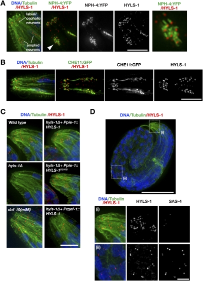
Figure 4.
HYLS-1 is present at the base of mature C. elegans cilia. (A) HYLS-1 localizes to the base of the cilium. Late (threefold)-stage embryo expressing the transition zone marker NPH-4:YFP stained for DNA, tubulin, and HYLS-1. The inset shows 3× magnified view of amphid bundle (arrowhead). (B) L1 larva expressing the IFT protein CHE-11:GFP stained for DNA, tubulin, and HYLS-1. (C) Expression of wild-type or disease mutant HYLS-1 from a germline promoter (Ppie-1) restores localization in hyls-1Δ animals, wheras expression from a pan-neuronal promoter (Prgef-1) fails to do so. Late-stage embryos stained for DNA, tubulin, and HYLS- 1. (D) SAS-4 is not present at the ciliary base (panel i), although it colocalizes with HYLS-1 at centrioles elsewhere in the animal (panel ii). Late-stage embryo stained for DNA, tubulin, HYLS-1, and SAS-4. Bars: A–C, 5 μm; D, top panel, 50 μm; D, panels i,ii, 10 μm.

