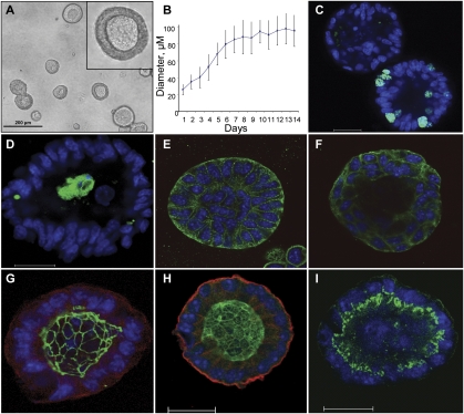
Figure 2.
Metastatic 344SQ cells form polarized epithelial spheres in 3D Matrigel culture. (A) Morphology of day 12 spheres by contrast microscopy. (B) Sphere growth versus time from plating onto Matrigel (MG); diameter measured in microns. (C) Ki-67 (green) and Topro-3 (blue) staining of developing sphere, visualized by confocal microscopy. (D) Cleaved caspase 3 staining (green) and Topro-3 (blue) of developing sphere. (E) β-catenin (green) and Topro-3 (blue) staining of developing sphere. (F) E-cadherin (green) and Topro-3 (blue) staining of developing sphere. (G) Sphere stained for ZO-1 (green), α6-integrin (red), and Topro-3 (blue). (H) Sphere stained for ParD6B (green), α6-integrin (red), and Topro-3 (blue). (I) Sphere stained for GM130 (green) and Topro-3 (blue). Bar in confocal images represents 20 μm.

