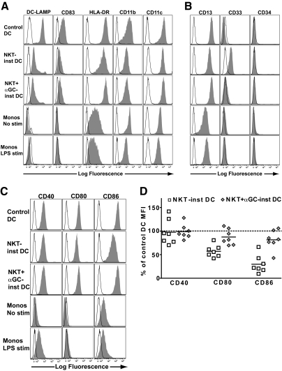
Figure 1.
Phenotype NKT-instructed APCs. Purified peripheral blood monocytes were cultured with recombinant GM-CSF and IL-4 (Control DC), or exposed to transwell inserts containing NKT cells with untreated or α-GalCer pulsed monocytes (NKT-inst APC or NKT+αGC-inst APC, respectively). After 3 days of differentiation, the antigen-presenting cells (APCs) were washed and then matured by exposure to LPS for an additional 48 h. As controls, monocytes cultured in medium alone for 2 days (Monos No stim), or monocytes treated with LPS for 2 days (Monos LPS stim), were also analyzed. (A) Flow cytometric analysis of expression of markers associated with mature myeloid dendritic cells (DCs). The APCs were stained with the indicated mAbs (solid histograms) or with isotype-matched negative control antibodies (open histograms). For analysis of DC-LAMP, the APCs were fixed and permeabilized prior to staining. One representative experiment out of 7 independent analyses is shown. (B) Analysis of expression of markers associated with myeloid-derived suppressor cells (MDSCs). Results are representative of 7 independent experiments. (C) Analysis of expression of costimulatory molecules. (D) Cell surface expression levels of the indicated costimulatory molecules on APCs that were exposed to NKT cells with unpulsed monocytes (white squares) or NKT cells with α-GalCer pulsed monocytes (gray diamonds), shown as a percentage of the corresponding marker on control DCs. Each symbol represents data from one independent experiment.

