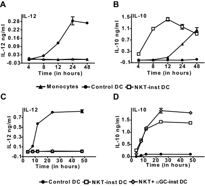
Figure 2.
APC cytokine production. Supernatants were withdrawn from cultures of control DCs, NKT-instructed APCs, or fresh monocytes at the indicated time points after the addition of LPS, and tested by standardized ELISAs for IL-12p70 (A) or IL-10 (B). (C and D) Comparison of APCs induced by auto-antigen or α-GalCer-activated NKT cells. Symbols and error bars show the means and standard deviations of 3 replicates (note that standard deviations are not always visible on the scales shown). Results are representative of 3-7 independent experiments. Similar results were obtained using ELISA reagents specific for IL-12p40 (data not shown).

