Figure 3.
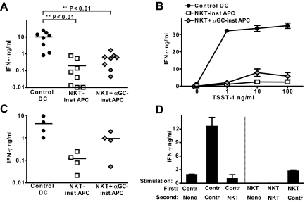
Stimulation of T cell IFN-γ production. APCs were prepared as described in Figure 1 and activated by exposure to LPS for 8 h. (A) Control DCs or NKT-instructed APCs were incubated with allogeneic peripheral blood T cells (i.e., total T cells), and supernatants were withdrawn after 24 h and analyzed for IFN-γ by ELISA. Each symbol corresponds to the amount of cytokine (shown on a log10 scale) from an independent allogeneic T cell + DC pairing. (B) Control DCs or NKT-instructed APCs were treated with the indicated concentrations of TSST-1 superantigen and used to stimulate IFN-γ secretion by autologous T cells. Symbols and error bars show the means and standard deviations of 3 replicates. The results are from one representative experiment out of 3 independent analyses. (C) APCs were incubated with allogeneic memory T cells (i.e., the CD45RO+ subset of peripheral blood T cells). (D) Stimulation of IFN-γ secretion by preactivated T cells. Allogeneic T cells were incubated for 3 days with control DCs or NKT-instructed APCs as a first stimulus. The T cells were harvested and then stimulated with the indicated APCs, and supernatants were collected after 24 h and analyzed for IFN-γ by ELISA. The plot shows the means and standard deviations of 3 replicate samples. Similar results were obtained in 3 independent experiments.

