Figure 5.
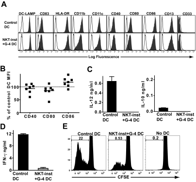
Dominant effect of NKT cell instruction. (A) Monocytes were cultured in medium containing recombinant GM-CSF and IL-4 alone (Control DC) or with recombinant GM-CSF and IL-4 in the presence of a transwell insert containing NKT cells and monocytes (NKT-inst + G-4 DC), then matured with LPS. The resulting cells were stained with the indicated mAbs (solid histograms) or with isotype-matched negative control mAbs (open histograms). The results are from one representative experiment out of 7 independent analyses. (B) Cell surface expression levels of the indicated costimulatory molecules on DCs that were cultured with GM-CSF and IL-4 in the presence of NKT cell factors, shown as a percentage of the corresponding marker on control DCs. Each symbol represents data from one independent experiment. (C) DCs were stimulated with LPS, and after 24-h culture supernatants were tested by ELISA for IL-12p70 and IL-10. Results are representative of 4 independent experiments. Similar results were obtained using ELISA reagents specific for IL-12p40 (data not shown). (D) Stimulation of IFN-γ secretion by allogeneic T cells. Results are from one representative experiment out of 4 independent analyses. (E) Stimulation of allogeneic T cell proliferation. The histograms show the CFSE intensity of the live T cells (i.e., DAPI−/CD3+ cells). Results shown are from one representative experiment out of 3 independent analyses.

