Figure 7.
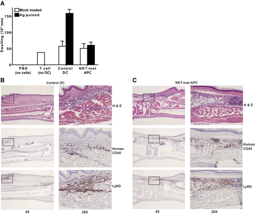
Noninflammatory phenotype of NKT-instructed APCs. (A) Control DCs or NKT-instructed APCs were pulsed with recall antigens (solid bars) or mock-treated (open bars) in the presence of LPS for 8 h, then mixed with purified autologous T cells and injected into the footpads of CB17 SCID mice, and the footpad swelling was measured after 24 h. Control footpads were injected with sterile PBS buffer (no cells) or with human T cells that were not mixed with APCs (no DC). The plot shows the means and standard deviations of the net swelling from 6-10 independent experiments. (B and C) Histological analysis of murine footpads that were injected with human T cells and antigen-pulsed control DCs (B) or human T cells and antigen-pulsed NKT-instructed APCs (C). Serial sections were stained with hematoxylin and eosin (top panels), or immunolabeled for human CD45 (middle panels), or for the murine neutrophil marker Ly6G (bottom panels). Panels on the left show longitudinal serial sections of footpad at ×4 magnification. The boxed areas of the left panels are shown at ×20 magnification on the right.

