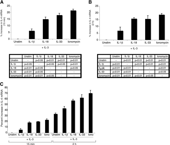
Figure 3.
IL-4 mRNA expression in cytokine-stimulated basophils. Basophils were stimulated with IL-3 (10 ng/mL) alone or in combination with IL-1β (10 ng/mL), IL-18 (20 ng/mL), IL-33 (10 ng/mL), or ionomycin (1 μm) for 15 min (A and C) or 2 h (B and C). IL-4 mRNA levels were analyzed along with β-actin as a housekeeping gene by semiquantitative PCR. IL-4 mRNA levels were normalized to β-actin and graphed as the percent increase relative to IL-3-only stimulation at 15 min (A and C) or IL-3-only stimulation at 2 h (B). A representative gel is shown in Supplemental Figure 4. Results are the mean of three experiments ± sem. Statistical significance data are reported for each time-point below the respective graph.

